What is Facet Syndrome? (Arthropy)
Facet arthropathy commonly occurs due to aging, wear and tear, injury, and some occupations are more prone to this. As the facet joint cartilage wears out it develops bone on bone rubbing which stimulates more bone and thicker bone to form. This is what causes bone spurs to form which may lead to stenosis and also possibly may result in instability called spondylolisthesis. This often causes not only back pain but also arm or leg pain and radicular symptoms.
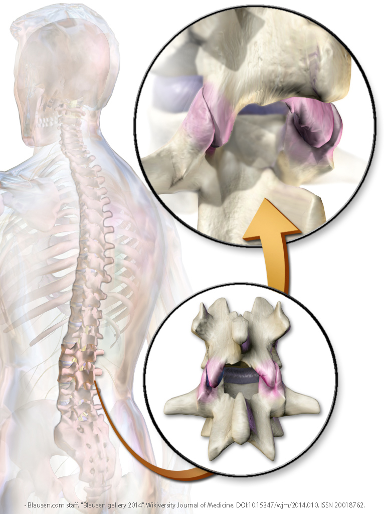 Blausen.com staff. "Blausen gallery 2014". Wikiversity Journal of Medicine. DOI:10.15347/wjm/2014.010. ISSN 20018762. - Own work
How is Facet Arthritis Diagnosed?
Facet arthropathy may be diagnosed on x-rays, CT and MRI. CT scan may show thickened irregular facet joints. MRI scan may show joint swelling, thickened ligaments and bones and pinched nerves. These imaging studies only show the physical abnormality. They do not indicate if these changes are responsible for your pain.
To determine the painful structure the patient may undergo pain mapping. Facet based injections are used to localize where the pain is coming from. A local anesthetic is injected into the facet joint or onto the facet nerve (medial branch nerve). If the pain is caused by the facet joint, the pain will stop or decrease. If the pain does not improve then the pain is not from the facet joint, but is originating from a different pain generator. This may include a different level other than the one tested, the disk space, or the sacroiliac joint.
What are the Treatment Options?
Patients who fail conservative treatment may benefit from surgical treatment. Traditionally these patients were treated with either a large fusion surgery or radiofrequency ablation (RFA). RFA is the placement of burning electrodes blindly onto the spine under x-ray guidance to burn medial branch nerves that transmit back pain. This method of pain relief lasts up to 18 months.
Endoscopic Rhizotomy is a new improved treatment for back pain. The medial branch nerves are found and cut under direct visualization through the endoscope. Patients can have immediate pain relief lasting for years. Recovery time ranges from one to three weeks.